Brillouin scattering (BS) spectroscopy is an inelastic light scattering technique, similar to the Raman spectroscopy. However, the energies of the excitations probed in the BS spectroscopy are much lower than those probed in the Raman spectroscopy and they lie well below 50 cm-1. The excitations that can be investigated with BS spectroscopy include acoustic phonons, magnons, spin waves, surface ripples, diffusive excitations, etc. Acoustic phonons. produce strain, causing the change of mass density, and modulate the dielectric constant of the material. The BS spectroscopy allows for the monitoring of elastooptic properties of the material, the determination of sound velocities and elastic constants, the investigation of the effects such as anharmonicity, the phase transitions. It also provides information on the interaction of acoustic phonons with different excitations, electronic states, polaritons, and plasmons. The spectra are usually recorded in the backscattering geometry since forward scattering can be performed only on transparent samples.
Since the energy of the scattered radiation is very close to the initial, laser line energy, differing from it often for less than an inverse centimeter, a grating monochromator cannot be used as a spectrograph in this experiment. Instead, a Fabry Perot interferometer is used (plane or spherical). The plane Fabry Perot interferometer consists of two parallel disc-shaped mirrors made of glass or quartz mounted at a constant distance L one from the other. The flat mirror surfaces are coated with such a material that most of the light incident upon them is reflected and only a small fraction is transmitted. The interference of light after numerous reflections has maxima for wavelengths: λ=2L/p (p is a non-zero integer) and by transmitting mostly light of these wavelengths, the individual wavelengths are separated from the rest of the scattered radiation.
Our Brillouin scattering system, the Tandem Fabry Perot Interferometer TF-1 (JRS Scientific), consists of two interferometers placed with their main axes being almost parallel, with a small offset angle φ that their mirror distances are almost the same, having values L and Lcosφ. The light is passed three times through each interferometer (six passes in total) and improves the contrast drastically. Two interferometers are piezoelectrically scanned and their work is synchronized by mounting their scanning mirrors on the common scanning stage. Also, by placing the interferometers in proximity to each other the system is less sensitive to temperature fluctuations. The mirror spacing can be set in the range (0-50) mm, whereas small mirror movements with scanning amplitude of up to 2 μm are made possible by use of deformable parallelogram beneath the mirrors holder and an attached transducer controlled electronically. Both interferometers are isolated from the low-frequency vibrations (such as building vibrations) with a vibration free system AVI 350 LP.
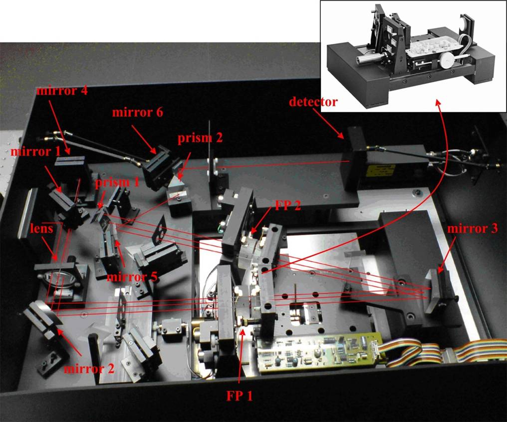
Brillouin JRS Scientific
spectrometer.
Inset: Tandem Fabry Perot interferometer with a common scanning
stage
A complete spectrometer is shown in the figure above. On the left side the beam enters the system through the aperture, then is reflected from the small mirror 1, collected and collimated with the lens, reflected from the mirror 2 and after this, it enters the Fabry Perot interferometer 1 (FP1). After a signal intensity amplification in the FP1, the beam is reflected from the mirror 3, passes through the second interferometer (FP2), is refracted and reflected from the prism 1 and returns through the FP2 and FP1 to the lens and is focused on the very small mirror 4 (cat’s eye) behind the first mirror. From the mirror 4, the beam returns again through the FP1 and FP2, but now is reflected from the mirror 5, refracted through the prism 2, reflected from the mirror 6 and through a small lens is directed to the detector. It is clear that the beam passes three times through a FP1 and three times through a FP2. The distance between the mirrors is set by using a control on the front board that runs a motor that moves the translation stage. The adjustment of the FP1 and FP2 mirrors positions is performed also with x- and y-position controls on the front board, with a purpose of making FP1 and FP2 transmit. A fine adjustment of these positions is performed with the help of corresponding controls on the control unit. The beam transmission is controlled visually, checking that the entire beam spot is viewed on a piece of paper placed after the FP1 and the FP2, after mirror 6 etc. The described procedure relates to the tandem operating mode of the spectrometer. There is one more working mode, the alignment mode, with a slightly changed geometry, which is characterized by the collection of the light reflected from the FP1 and the FP2, which allows for viewing dips at the positions in the spectra corresponding to the wavelengths transmitted through the interferometers. This operation mode makes it easier to do final adjustments of mirror positions in order to ensure that the interferometers transmit completely. After this a measurement is made by setting the scanning amplitude on the control unit and collecting the spectra. The signal from the detector (photon counting module Perkin Elmer MP 983) is analyzed with a MCA Ghost multichannel analyzer.
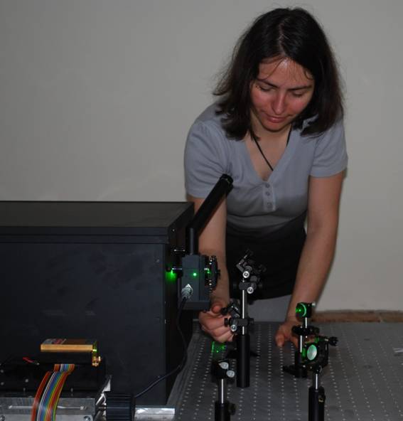
Brillouin
scattering experiment
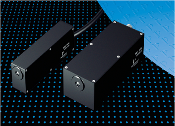
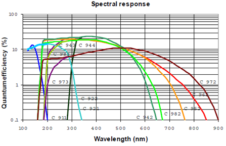
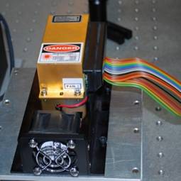
Detector incorporated in the experiment is PerkinElmer Photon Counting
Module MP 983, with spectral sensitivity in the region (185-650) nm.
Photocatode used is a low noise bialkali, with dark counts of 2cps, and
maximum counting rate of 10MHz. Detector and the spectral response of
the photocatode (curve signed C983) is shown in the figure down.
 |
 |
 |
|
|
Photon Counting Module MP 983 (left), with spectral sensitivity–red curve C983 (right). |
Coherent Compass 315 M laser. |
||
Excitation source in the BS spectroscopy experiment is a Coherent diode-pumped Nd:YAG laser, model Compass 315M. It is a single frequency, single mode laser operating at the wavelength of 532 nm with maximum power of 150mW.
Center
for Solid State and New Materials :: Facilities
:: Laboratory for
Brillouin scattering spectroscopy
::
print